45 microscope with labels and functions
› fluorescent-dyesFluorescent Dyes Types, Vs Proteins, Applications Etc. Fluorescent dyes are divided into several groups that include: Organic dyes; Biological fluorophores Organic Fluorescent Dyes. Essentially, organic dyes are characterized by emissions originating from optical transition delocalized over the whole chromophore or from intermolecular charge transfer transitions (intramolecular charge transfer from the excited electronic state). Lipid bilayer - Wikipedia The lipid bilayer is very thin compared to its lateral dimensions. If a typical mammalian cell (diameter ~10 micrometers) were magnified to the size of a watermelon (~1 ft/30 cm), the lipid bilayer making up the plasma membrane would be about as thick as a piece of office paper. Despite being only a few nanometers thick, the bilayer is composed of several distinct chemical …
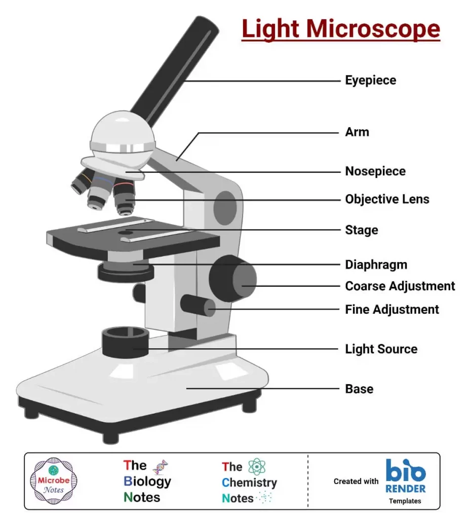
Parts of a microscope with functions and labeled diagram - Microbe Notes Microscopes are instruments that are used in science laboratories to visualize very minute objects such as cells, and microorganisms, giving a contrasting image that is magnified. Microscopes are made up of lenses for magnification, each with its own magnification powers.
Microscope with labels and functions
Label the microscope — Science Learning Hub All microscopes share features in common. In this interactive, you can label the different parts of a microscope. Use this with the Microscope parts activity to help students identify and label the main parts of a microscope and then describe their functions. Drag and drop the text labels onto the microscope diagram. A Study of the Microscope and its Functions With a Labeled Diagram ... A Study of the Microscope and its Functions With a Labeled Diagram To better understand the structure and function of a microscope, we need to take a look at the labeled microscope diagrams of the compound and electron microscope. These diagrams clearly explain the functioning of the microscopes along with their respective parts. Microscope Types (with labeled diagrams) and Functions Simple microscope labeled diagram Simple microscope functions It is used in industrial applications like: Watchmakers to assemble watches Cloth industry to count the number of threads or fibers in a cloth Jewelers to examine the finer parts of jewelry Miniature artists to examine and build their work Also used to inspect finer details on products
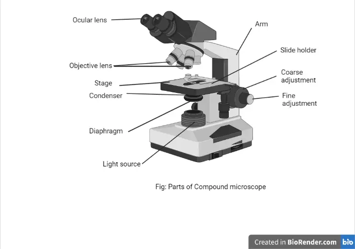
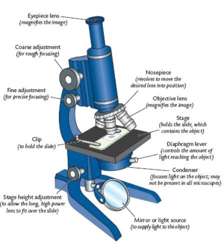
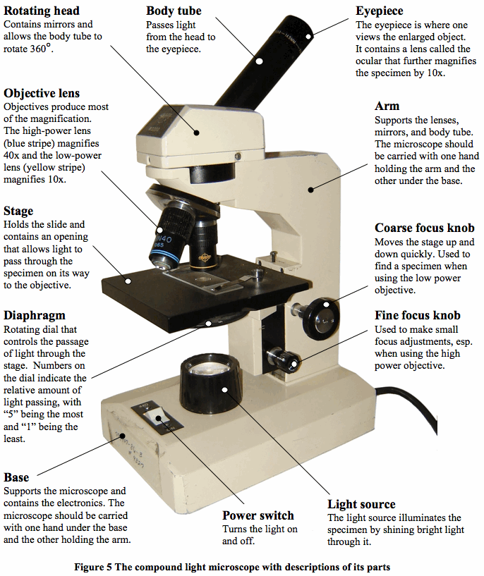
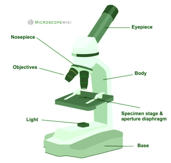
Microscope with labels and functions. Parts of the Microscope with Labeling (also Free Printouts) Let us take a look at the different parts of microscopes and their respective functions. 1. Eyepiece it is the topmost part of the microscope. Through the eyepiece, you can visualize the object being studied. Its magnification capacity ranges between 10 and 15 times. 2. Body tube/Head It is the structure that connects the eyepiece to the lenses. Compound Microscope Parts, Functions, and Labeled Diagram Compound Microscope Parts, Functions, and Labeled Diagram Parts of a Compound Microscope Each part of the compound microscope serves its own unique function, with each being important to the function of the scope as a whole. rsscience.com › stereo-microscopeParts of Stereo Microscope (Dissecting microscope) – labeled ... Unlike a compound microscope that offers a flat image, stereo microscopes give the viewer a 3-dimensional image that you can see the texture of a larger specimen. [In this image] Examples of Stereo & Dissecting microscopes. Major microscope brands (Zeiss, Olympus, Nikon, Amscope, Omano, Leica …) all produce stereomicroscopes. Microscope Parts, Function, & Labeled Diagram - slidingmotion Microscope parts labeled diagram gives us all the information about its parts and their position in the microscope. Microscope Parts Labeled Diagram The principle of the Microscope gives you an exact reason to use it. It works on the 3 principles. Magnification Resolving Power Numerical Aperture. Parts of Microscope Head Base Arm Eyepiece Lens
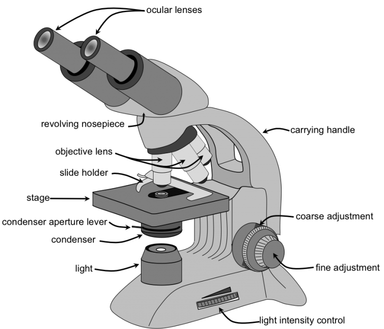
Compound Microscope Parts, Functions, and Labeled Diagram Nov 18, 2020 · Parts of a Compound Microscope Each part of thenbsp compound microscope serves its own unique function, with each being important to the function of the scope as a whole. The individual parts of a compound microscope can vary heavily depending on the configuration & applications that the scope is being used for. Common compound microscope parts include: … Microscope, Microscope Parts, Labeled Diagram, and Functions (2022) Microscope, Microscope Parts, Labeled Diagram, and Functions (2022) Table of Contents. Structure of ... Types of Microscope FAQ About Microscope and Microscope Parts. A microscope is a laboratory instrument used to examine objects that are too small to be seen by the naked eye. It is derived from Ancient Greek words and composed of mikrós ... Binocular Microscope Anatomy - Parts and Functions with a Labeled ... Ocular lens or eyepiece of the microscope, Diopter adjustment of the eyepiece All of these parts are identified in a light microscope labeled diagram. So, first, make sure you can identify all these parts from this labeled diagram. Parts of the compound microscope › dissecting-stereoDissecting Stereo Microscope Parts and Functions Dissecting Stereo Microscope Parts and Functions Overview. Also known as a stereoscopic microscope, a dissecting microscope is a type of optical microscope commonly used for studying three-dimensional objects (3-D objects) as well as for dissecting biological specimen (e.g. insects and plant parts etc) at low magnification, between 2 and 100x depending on the microscope.
en.wikipedia.org › wiki › Lipid_bilayerLipid bilayer - Wikipedia Lipid bilayers cannot be seen in a traditional microscope because they are too thin. In order to see bilayers, researchers often use fluorescence microscopy . A sample is excited with one wavelength of light and observed in a different wavelength, so that only fluorescent molecules with a matching excitation and emission profile will be seen. Shop by Category | eBay Shop by department, purchase cars, fashion apparel, collectibles, sporting goods, cameras, baby items, and everything else on eBay, the world's online marketplace Microscope Parts and Functions First, the purpose of a microscope is to magnify a small object or to magnify the fine details of a larger object in order to examine minute specimens that cannot be seen by the naked eye. Here are the important compound microscope parts... Eyepiece: The lens the viewer looks through to see the specimen. Microscope, Microscope Parts, Labeled Diagram, and Functions Illuminator: Illuminator is the most important microscope parts and it serve as light source for a microscope during slide specimen visualization. It is a continuous source of light (110 volts) used in place of a mirror. The mirror of microscope is used to reflect light from the external light source up through the bottom of the stage.
Parts of a Microscope with Their Functions – Microbe Online
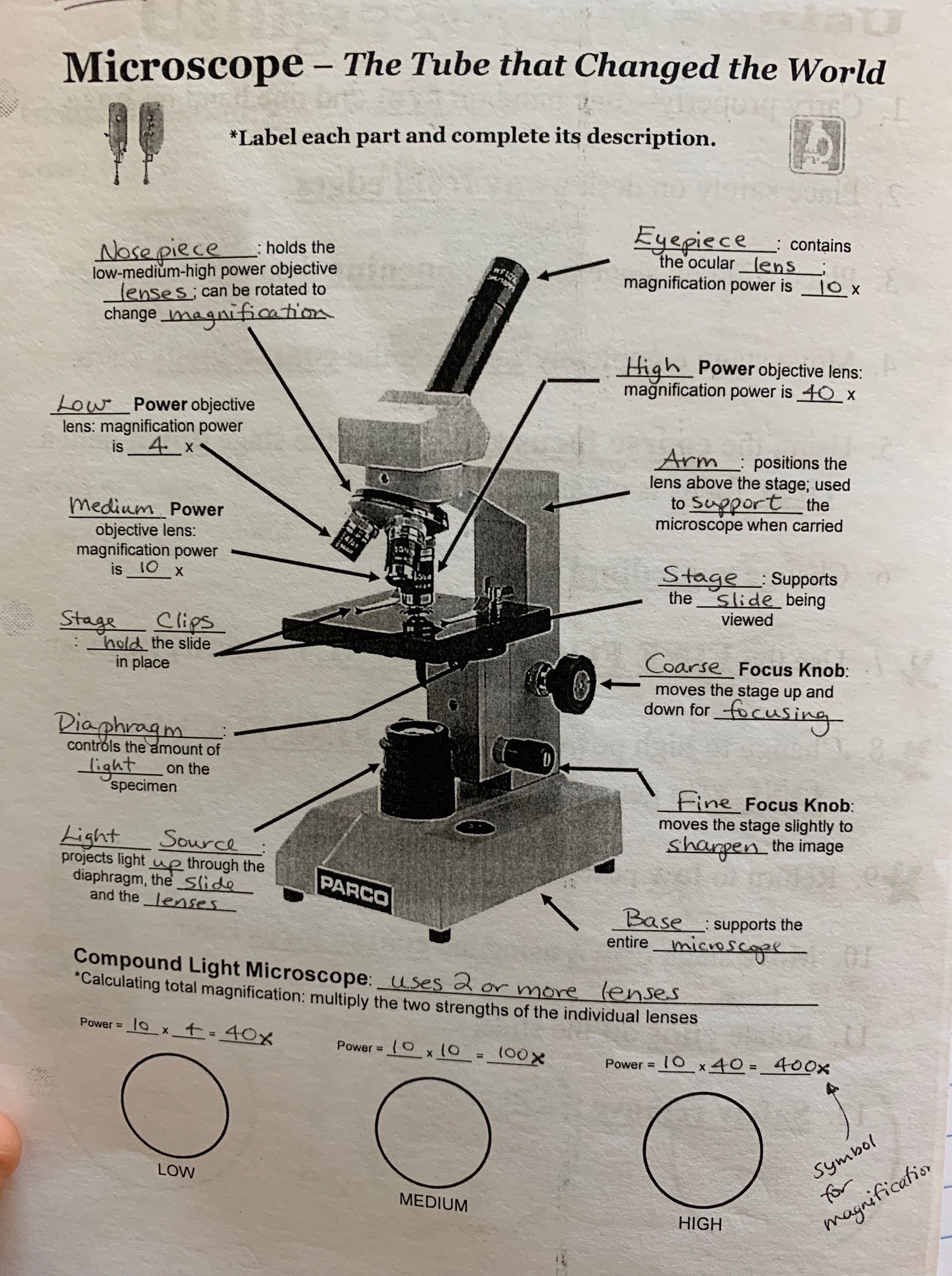
Compound Microscope Parts - Labeled Diagram and their Functions There are two major optical lens parts of a microscope: Eyepiece (10x) and Objective lenses (4x, 10x, 40x, 100x). Total magnification power is calculated by multiplying the magnification of the eyepiece and objective lens. The illuminator provides a source of light. The light is focused by the condenser and passing through the specimen placed ...
22 Parts Of a Microscope With Their Function And Labeled ...
Microscope With Labeled Parts and Functions - 24 Hours Of Biology Optical parts and the functions The optical parts of the microscope are used to view, enlarge, and produce an image from a sample placed on a slide. These parts include Eyepiece: Eyepiece also contains ocular lens. It enhance the image of the viewer. This part is used for checking through the microscope. Eyepiece is found at the upper part of it.

Microscope Parts and Functions - YouTube
Simple Microscope - Parts, Functions, Diagram and Labelling Stereo microscope/dissecting microscope - It can magnify objects by up to 300 times. It is used to visualize opaque objects that cannot be visualized using a compound microscope. Confocal microscope - It uses laser light to scan a dyed sample. Scanning electron microscope - Instead of light, this type of microscope uses electron.

Light Microscope: Parts and Function - ppt download
Fluorescent Dyes Types, Vs Proteins, Applications Etc. Fluorescent Dyes Types, Vs Proteins, Applications Etc. Overview. Fluorescent dyes (also known as fluorophores/reactive dyes) may simply be described as molecules (non-protein in nature) that, in microscopy, achieve their function by absorbing light at a given wavelength and re-emitting it at a longer wavelength.

What is a Compound Microscope? | Microscope World Blog
en.wikipedia.org › wiki › Electron_microscopeElectron microscope - Wikipedia An electron microscope is a microscope that uses a beam of accelerated electrons as a source of illumination. As the wavelength of an electron can be up to 100,000 times shorter than that of visible light photons , electron microscopes have a higher resolving power than light microscopes and can reveal the structure of smaller objects.
Free Microscope Drawing, Download Free Microscope Drawing png ...
en.wikipedia.org › wiki › Two-photon_excitationTwo-photon excitation microscopy - Wikipedia In scattering tissue, on the other hand, the superior optical sectioning and light detection capabilities of the two-photon microscope result in better performance. Applications Main. Two-photon microscopy has been involved with numerous fields including: physiology, neurobiology, embryology and tissue engineering.

Compound Microscope Parts – Labeled Diagram and their ...
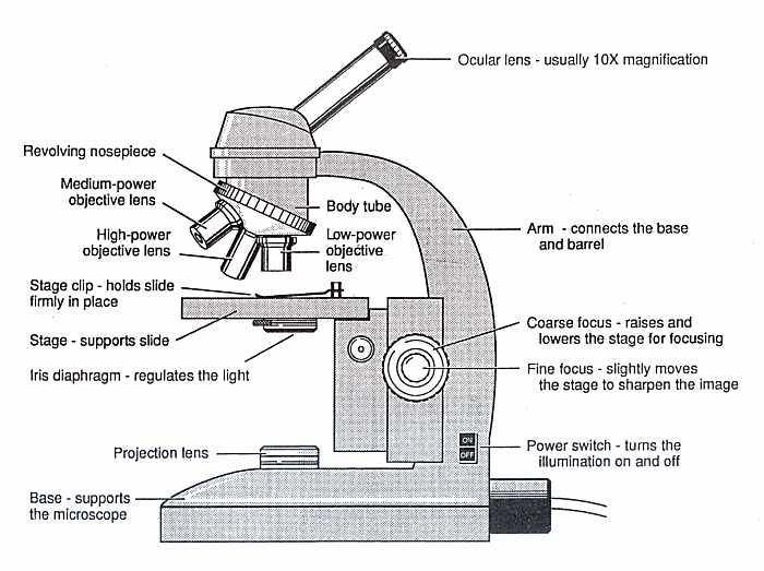
Microscope labeling and functions Flashcards | Quizlet Coarse Adjustment Knob Moves the body tube slightly to adjust the image; Fine Adjustment Knob Supports the body tube Arm Supports the slide being used Stage Projects or reflects light upward through the diaphragm Light Source Supports the microscope Base Body tube What is #1 called? Revolving nosepiece What is #2 called? Sets with similar terms
Understanding the Compound Microscope Parts and its Functions ...
Cross-tissue immune cell analysis reveals tissue-specific ... - Science Here we present an immune cell atlas of myeloid and lymphoid lineages across adult human tissues. We developed CellTypist for automated immune cell annotation and performed an in-depth dissection of cell populations, identifying 101 cell types or states from more than one million cells, including previously underappreciated cell states.

microscope | Types, Parts, History, Diagram, & Facts | Britannica
Electron microscope - Wikipedia An electron microscope is a microscope that uses a beam of accelerated electrons as a source of illumination. As the wavelength of an electron can be up to 100,000 times shorter than that of visible light photons, electron microscopes have a higher resolving power than light microscopes and can reveal the structure of smaller objects. A scanning transmission electron microscope …

Compound Microscope Parts – Labeled Diagram and their ...
Parts of Stereo Microscope (Dissecting microscope) – labeled … Unlike a compound microscope that offers a flat image, stereo microscopes give the viewer a 3-dimensional image that you can see the texture of a larger specimen. [In this image] Examples of Stereo & Dissecting microscopes. Major microscope brands (Zeiss, Olympus, Nikon, Amscope, Omano, Leica …) all produce stereomicroscopes.
Parts of a Compound Microscope (And their Functions)
Microscope: Parts Of A Microscope With Functions And Labeled Diagram. Q. Define a Microscope. Ans. Microscopes are instruments that are used in science laboratories, to visualize very minute objects such as cells, and microorganisms, giving a contrasting image, that is magnified. Q. State functions of a microscope. Ans. A microscope is usually used for the study of microscopic algae, fungi, and biological specimens.

Microscope labeling and functions Flashcards | Quizlet
Microscope Labels and Functions Flashcards | Quizlet Microscope Labels and Functions. STUDY. PLAY. Ocular Lenses (eyepiece) magnifies. Arm. supports the tube and connects it to the base. Revolving nosepiece. holds the objective lenses and can be rotated to easily change the power. Objective lenses. magnifies. Coarse adjustment knob.
Compound and Stereo- microscopes - Microscopes 4 Schools
Two-photon excitation microscopy - Wikipedia Two-photon excitation microscopy (TPEF or 2PEF) is a fluorescence imaging technique that allows imaging of living tissue up to about one millimeter in thickness, with 0.64 μm lateral and 3.35 μm axial spatial resolution. Unlike traditional fluorescence microscopy, in which the excitation wavelength is shorter than the emission wavelength, two-photon excitation requires …

The Different Types and Uses of a Stereo Microscope -
Microscope Parts & Functions - AmScope Main Microscope Parts and Functions. How to Identify Parts on a Compound Microscope. Watch on. Head: The upper part of the microscope houses the eyepiece and objective lenses. Tube: Where the eyepieces are dropped in. Also, it connects the eyepieces to the objective lenses. Stage: The flat platform that supports the slides.

Compound Microscope Educational Resources K12 Learning, Life ...
› liver_disease › articleLiver Disease: Early Signs, Symptoms, Treatment, Stages ... Mar 23, 2022 · The liver has many functions. Signs and symptoms of liver disease include abdominal pain, jaundice, nausea, and weakness. Causes, treatment, and life expectancy vary. Lifestyle changes may slow the progression of some types of liver disease.

Compound Microscope Parts, Functions, and Labeled Diagram ...
Microscope labels and functions Flashcards | Quizlet stage clips holds the slide in place Diaphragm controls the amount of light entering coarse adjustment knob makes large adjustments (moves stage up and down quickly) fine adjustment knob makes small adjustments (slightly moves stage to improve focus) base supports the microscope arm supports microscope magnification Sets with similar terms

Types, Parts and Functions of a Microscope
Microscope Parts and Functions Flashcards | Quizlet Magnifies image 40X found on the nosepiece. Base. Support/bottom of the microscope, used to carry the microscope. Light Source. Provides light to enable us to see the specimen on the slide. Arm. Used in order to carry the microscope. Coarse Adjustment. moves the stage up or down a lot, used first when viewing the slide.
(159).jpg)
Microscope Quiz: How Much You Know About Microscope Parts And ...
Dissecting Stereo Microscope Parts and Functions Dissecting Stereo Microscope Parts and Functions Overview. Also known as a stereoscopic microscope, a dissecting microscope is a type of optical microscope commonly used for studying three-dimensional objects (3-D objects) as well as for dissecting biological specimen (e.g. insects and plant parts etc) at low magnification, between 2 and 100x depending on the …
compound microscope with label and functions - Clip Art Library
Liver Disease: Early Signs, Symptoms, Treatment, Stages, Types Mar 23, 2022 · This is the reason that warning labels exist on many over-the-counter medications that contain acetaminophen and why prescription narcotic-acetaminophen combination medications (for example, Vicodin, Lortab, Norco, Tylenol #3) limit the numbers of tablets to be taken in a day. For patients with underlying liver disease or those who abuse ...

Compound Microscope: Parts of Compound Microscope
Microscope Types (with labeled diagrams) and Functions Simple microscope labeled diagram Simple microscope functions It is used in industrial applications like: Watchmakers to assemble watches Cloth industry to count the number of threads or fibers in a cloth Jewelers to examine the finer parts of jewelry Miniature artists to examine and build their work Also used to inspect finer details on products
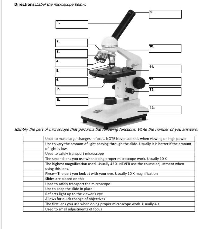
Solved Directions:Label the microscope below. 9. 1. 111 2 ...
A Study of the Microscope and its Functions With a Labeled Diagram ... A Study of the Microscope and its Functions With a Labeled Diagram To better understand the structure and function of a microscope, we need to take a look at the labeled microscope diagrams of the compound and electron microscope. These diagrams clearly explain the functioning of the microscopes along with their respective parts.

Microscope Diagram Labeled, Unlabeled and Blank | Parts of a ...
Label the microscope — Science Learning Hub All microscopes share features in common. In this interactive, you can label the different parts of a microscope. Use this with the Microscope parts activity to help students identify and label the main parts of a microscope and then describe their functions. Drag and drop the text labels onto the microscope diagram.

Compound Microscope Parts – Labeled Diagram and their ...
16 Types of Microscopes with Parts, Functions, Diagrams
Understanding the Compound Microscope Parts and its Functions ...

Parts of a Microscope Labeling Activity

Compound Microscope Parts and Functions | Science fair ...

Compound Microscope Parts, Diagram Definition, Application ...
Compound Microscope Parts, Functions, and Labeled Diagram ...

Microscope With Labels clip art | Microscope parts ...

Label the microscope — Science Learning Hub

What Are Parts Of Microscope And Their Function? - Fun Biology
Microscope Parts - Ms. Ang's Class

Biology Microscope Parts and Functions Diagram | Quizlet
compound microscope with label and functions - Clip Art Library

Label the microscope — Science Learning Hub

Pin on Micellaneous

13 parts of the Compound Light Microscope Diagram | Quizlet
microscope parts and functions - Quizizz

Simple Microscope - Parts, Functions, Diagram and Labelling ...
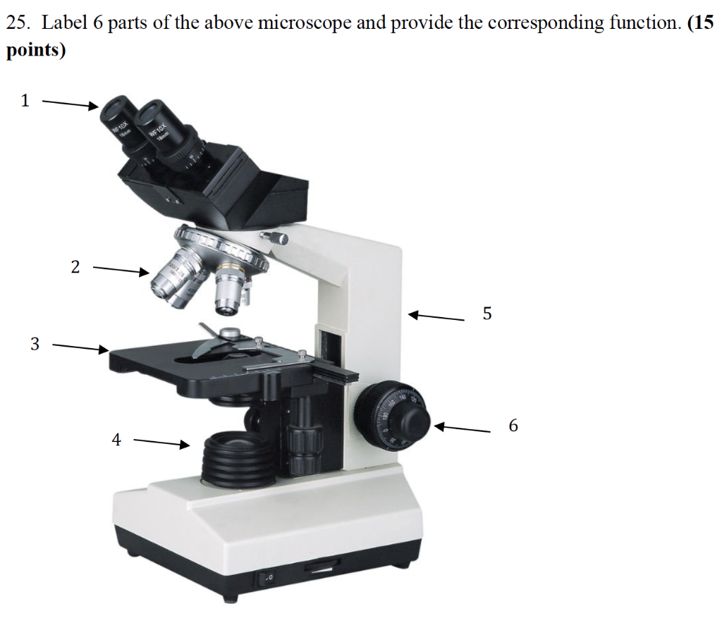
Solved 25. Label 6 parts of the above microscope and provide ...
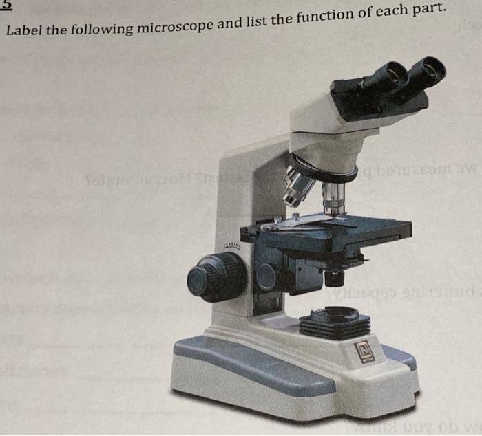
Solved Label the following microscope and list the function ...

Compound Microscope Drawing With Parts and Functions
Microscope Types (with labeled diagrams) and Functions

Types, Parts and Functions of a Microscope
Post a Comment for "45 microscope with labels and functions"