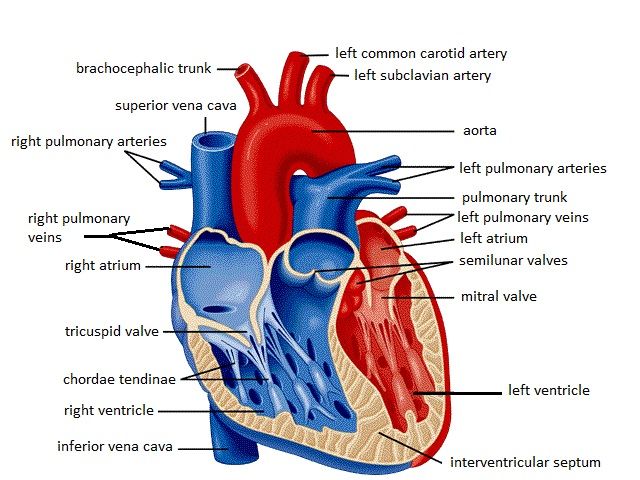
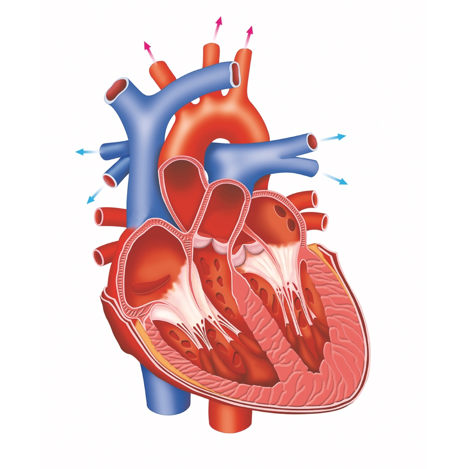
44 diagram of a human heart with labels
Layers of the heart: Epicardium, myocardium, endocardium - Kenhub Heart Cor 1/4 This article will discuss the layers of the heart (the epicardium, the myocardium and the endocardium) and any clinical relations pertaining to them. In the same way that vehicles have their fuel pumps, our body has the heart. The heart is a muscular organ found in the middle mediastinum that pumps blood throughout the body. Anatomy of the heart and coronary arteries (coronary CT) - IMAIOS ISSN 2534-5079 Anatomical parts 1. Basal anterior 10. Mid inferior 11. Mid inferolateral 12. Mid anterolateral 13. Apical anterior 14. Apical septal 15. Apical inferior 16. Apical lateral 17. Apex 2. Basal anteroseptal 3. Basal inferoseptal 4. Basal inferior 5. Basal inferolateral 6. Basal anterolateral 7. Mid anterior 8. Mid anteroseptal 9.
Basic Human Heart Diagram Labeled - Simple Human Lungs And Heart ... Diagram of circulatory system with main parts labeled. Find the perfect human heart diagram stock photo. It may be a straight tube, as in spiders and annelid worms, or a somewhat more elaborate structure with one or more receiving chambers (atria) and a main . Human heart beats over 100,000 times a day?

Diagram of a human heart with labels
The Ultimate Heart Model & Sheep Heart Practice Quiz! - ProProfs Take this practice quiz that covers information related to the sheep heart & the heart model. It is intended for use as a supplemental study aid. As is the case in the lab practical, each correct answer counts. So, make sure you learn from the feedback. Questions and Answers. 1. Labeled diagram of the heart along with the various ... - Kelsi Schanbacher Heart diagram of coronary artery function . Labeled diagram of the heart along with the various blood vessels and valves. 9 marks veins capillaries arteries heart blood vessel plasma platelets. It's just really crazy to see a diagram, but then actually do the . Grade 9 science students dissected pig hearts to understand the inner. Heart (right and left atrium): Anatomy and function - Kenhub Revise the anatomy of the atria and the other parts of the heart with our heart diagrams, quizzes and labeled worksheets. Once ventricular contraction stops and the pressure within the atria overcomes the pressure within the ventricles, the atrioventricular valves open and the blood passes into the ventricles.
Diagram of a human heart with labels. Heart | Radiology Reference Article | Radiopaedia.org Gross anatomy. The heart has a somewhat conical form and is enclosed by the pericardium. It is positioned posteriorly to the body of the sternum with one-third situated on the right and two-thirds on the left of the midline. Its left-sided orientation is formally known as levocardia (cf. dextrocardia ). Histology, Heart - StatPearls - NCBI Bookshelf The heart is a four-chambered organ responsible for pumping throughout the body. It receives deoxygenated blood from the body, sends it to the lung, receives oxygenated blood from the lungs, and then distributes the oxygenated blood throughout the body. At the histological level, the cellular features of the heart play a vital role in the normal function and adaptations of the heart. Body Cavities and Membranes: Labeled Diagram, Definitions Body cavity labeled diagram of organs they contain, membranes, and lateral views. Body cavity definitions and subdivisions in tables and charts. Ventral, dorsal, cranial, spinal, vertebral, thoracic, pleural, pericardial, mediastinum, abdominopelvic, abdominal, and pelvic cavities explained. Quiz yourself with the labeled views! Human heart: Anatomy, function & facts | Live Science The human heart is an organ that pumps blood throughout the body via the vessels of the circulatory system, supplying oxygen and nutrients to the tissues and removing carbon dioxide and other ...
Diagrams, quizzes and worksheets of the heart | Kenhub Labeled heart diagrams Take a look at our labeled heart diagrams (see below) to get an overview of all of the parts of the heart. Once you're feeling confident, you can test yourself using the unlabeled diagrams of the parts of the heart below. Labeled heart diagram showing the heart from anterior Unlabeled heart diagrams (free download!) Circulatory System Diagram - New Health Advisor Heart consists of four chambers namely right atria, left atria, right ventricle and left ventricle. Both the atrium and ventricles are separated from each other with a muscular septum. Valves are present in between atria and ventricle which helps in draining the blood from upper part to lower part of the body. Diagram of Human Heart and Blood Circulation in It Exterior of the Human Heart A heart diagram labeled will provide plenty of information about the structure of your heart, including the wall of your heart. The wall of the heart has three different layers, such as the Myocardium, the Epicardium, and the Endocardium. Here's more about these three layers. Epicardium Heart anatomy: Structure, valves, coronary vessels - Kenhub Heart anatomy. The heart has five surfaces: base (posterior), diaphragmatic (inferior), sternocostal (anterior), and left and right pulmonary surfaces. It also has several margins: right, left, superior, and inferior: The right margin is the small section of the right atrium that extends between the superior and inferior vena cava .
How the Heart Works: Diagram, Anatomy, Blood Flow The heart is located under the rib cage -- 2/3 of it is to the left of your breastbone (sternum) -- and between your lungs and above the diaphragm. The heart is about the size of a closed fist, weighs about 10.5 ounces, and is somewhat cone-shaped. It is covered by a sack termed the pericardium or pericardial sack. Human Circulatory System | Parts, Functions, Types Human Heart with parts label. The circulatory system is a network that carries blood throughout the body. The human circulatory system supplies the food and oxygen that is necessary for the body to survive. At the same time, it carries carbon dioxide and other waste materials away from ... Anatomy of the Heart: Blood Flow and Parts - Study.com The Heart. In your body, blood flows within a closed circuit of blood vessels. Blood is able to circulate around your body thanks to a muscular pump known as your heart. As we previously learned ... Heart: illustrated anatomy - e-Anatomy - IMAIOS ISSN 2534-5079 Anatomical parts 1 - RCA proximal 1. Basal anterior 10 - Second diagonal 10. Mid inferior 10a - Second diagonal a 11 - Proximal circumflex 11. Mid inferolateral 12 - Intermediate/anterolateral 12. Mid anterolateral 12a - Obtuse marginal a 12b - Obtuse marginal b 13 - Distal circumflex 13. Apical anterior 14 - Left posterolateral 14.
Heart anatomy - labeled | Human anatomy and physiology, Arteries anatomy, Heart anatomy
Heart Ventricles | Left & Right Ventricle Function | Study.com Human Heart Diagram and Function. The human heart is part of the cardiovascular system. The main function of the heart is to supply oxygenated blood to the rest of the body for survival and to thrive.
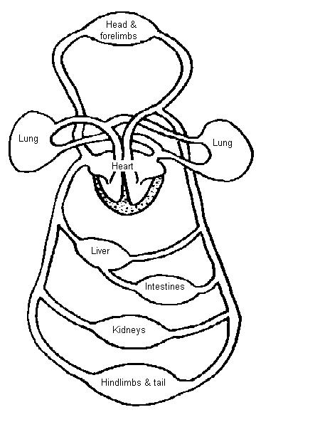
The Anatomy and Physiology of Animals/Circulatory System Worksheet - WikiEducator
Identify Various Parts Of A Human Heart: Trivia Quiz - ProProfs The heart is the most important organ in the body. It is in charge of keeping the processes within the body moving by facilitating the transfer of blood throughout the body. The quiz below is to test out interesting facts you may know about the heart. Give it a try and good luck.

Gaseous exchange in the lungs | Circulatory and respiratory systems | Siyavula
Internal Structure Of The Heart Labeled / Vector Illustration Diagram ... You can also look at the labeled pictures to get an idea of what the. 01.02.2014 · insulin plays a central role in the regulation of human metabolism. Internal Structure Of The Heart Labeled / Vector Illustration Diagram Human Heart Anatomy Stock Vector Royalty Free 177791534. You can also look at the labeled pictures to get an idea of what the.
labelled diagram of heart a level - Clip Art Library
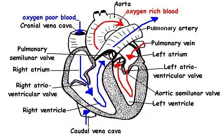
How the Heart Works - The Heart | NHLBI, NIH Blood also carries carbon dioxide to your lungs so you can breathe it out. Inside your heart, valves keep blood flowing in the right direction. Your heart's electrical system controls the rate and rhythm of your heartbeat. A healthy heart supplies your body with the right amount of blood at the rate needed to work well.

A Labeled Diagram of the Human Heart You Really Need to See | Human heart and Nursing students
Free Heart Worksheets for Human Anatomy Lessons Print out sheet of the human heart with labels - This fun heart worksheet shows kids the different parts of the heart. They'll learn about the left ventricle, the left atrium, the tricuspid valve, and more. Human Heart Clipart - There is a coloring page, heart labeling worksheet and heart anatomy chart.
heart labelled diagram | Diabetes Inc.
Know Where Your Heart Is and How to Identify Heart Pain Here we are going to discuss the symptoms of several chest pains which are associated with heart. 1. Heart Attack. Heart attack results from the occluded blood vessels that carry blood to the heart. The patient may experience the following signs: Fullness or squeezing sensation in the chest.

Leadership: Group Work Can Be As Successful As You Want It To Be
Human Heart for Kids: 2 Fun Heart Models plus Worksheets We used the parts of the heart diagram (in the pack below) to guide us. You may want to use a toothpick and paper to label the parts or play a fun review game to recall which part is which. Our hearts are divided into two parts - right and left halves. Each half is divided into two hollow sections called chambers.
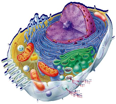
Free Animal Cell Unlabeled, Download Free Clip Art, Free Clip Art on Clipart Library
Heart Drawing With Label : Human Heart Diagram Images Stock Photos ... Most frequent question in exam to draw human heart diagram with labels. Png, jpg, gif · no labels version · azərbaycanca · català · english · english · hrvatski · italiano · lingua franca nova. Internal structure of human heart shows four chambers viz. Cell structure and functions / animal cell vs plant cell / parts of cell / ch 8 ...
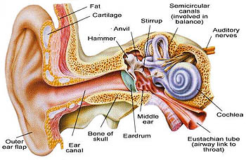
Ear Diagram - Science Photo (40502721) - Fanpop
Heart Labeling Quiz: How Much You Know About Heart Labeling? Create your own Quiz Here is a Heart labeling quiz for you. The human heart is a vital organ for every human. The more healthy your heart is, the longer the chances you have of surviving, so you better take care of it. Take the following quiz to know how much you know about your heart. Questions and Answers 1. What is #1? 2. What is #2? 3.

Detailed Labeled Anatomy Human Body | jpg: labeled heart flow | Nursing | Pinterest | Heart ...
Heart histology: Cells and layers - Kenhub The heart is a muscular, four-chambered system that is responsible for pumping blood through the vascular network. The organ is located within the thoracic cavity in a region known as the mediastinum. It is bordered bilaterally by the lungs, anteriorly by the sternum and posteriorly by the oesophagus and thoracic vertebra. Chambers

Human&Animal Anatomy and Physiology Diagrams: Heart and Great Vessels Diagram
Heart (right and left atrium): Anatomy and function - Kenhub Revise the anatomy of the atria and the other parts of the heart with our heart diagrams, quizzes and labeled worksheets. Once ventricular contraction stops and the pressure within the atria overcomes the pressure within the ventricles, the atrioventricular valves open and the blood passes into the ventricles.

Detailed Labeled Anatomy Human Body | jpg: labeled heart flow | Nursing | Pinterest | Beautiful ...
Labeled diagram of the heart along with the various ... - Kelsi Schanbacher Heart diagram of coronary artery function . Labeled diagram of the heart along with the various blood vessels and valves. 9 marks veins capillaries arteries heart blood vessel plasma platelets. It's just really crazy to see a diagram, but then actually do the . Grade 9 science students dissected pig hearts to understand the inner.

Human Heart Diagram
The Ultimate Heart Model & Sheep Heart Practice Quiz! - ProProfs Take this practice quiz that covers information related to the sheep heart & the heart model. It is intended for use as a supplemental study aid. As is the case in the lab practical, each correct answer counts. So, make sure you learn from the feedback. Questions and Answers. 1.
The Human Body: How does the heart work? – How It Works

The Heart Diagram Unlabeled | Projects to Try | Pinterest | Heart, The o'jays and Heart diagram

Pictures Of The Heart With Labels - koibana.info | Heart diagram, Human heart diagram, Human heart
Post a Comment for "44 diagram of a human heart with labels"