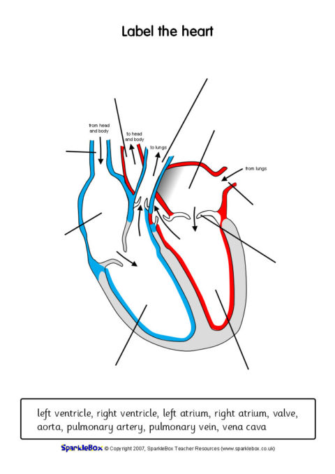
45 heart structure and labels
The Cardiovascular System Worksheet - Balancing Equations Worksheet Has a right side that pumps deoxygenated blood into the lungs to pick up oxygen. 2The upper part of the heart. Your heart pumps blood to every nook and cranny in your body. Comprised of the heart blood vessels and the blood itself it is divided into two loops which both begin in the heart. Rituximab - StatPearls - NCBI Bookshelf Rituximab, an anti CD20 monoclonal antibody, is used in the management of lymphoproliferative and autoimmune conditions. This activity reviews the indications, mechanism of action, contraindications, adverse effects, and other key factors (e.g., off-label uses, monitoring) pertinent for the healthcare team members.
Phenylephrine - StatPearls - NCBI Bookshelf Phenylephrine is less commonly used off-label as an additive to neuraxial/peripheral nerve blockade, to treat priapism and other conditions where local vasoconstrictive effects and reduced blood flow are the desired effect. ... Its chemical structure is related to epinephrine and ephedrine and possesses potent vasoconstriction properties when ...

Heart structure and labels
Histology, Lung - StatPearls - NCBI Bookshelf The respiratory portion of the lung consists of respiratory bronchiole, alveolar duct, alveolar sac, and finally alveoli where actual respiration takes place. In the conducting zone, the air is moistened, warmed, and filtered before it reaches the start of the respiratory region at the respiratory bronchioles. Circulatory system - Wikipedia The circulatory system includes the heart, blood vessels, and blood. The cardiovascular system in all vertebrates, consists of the heart and blood vessels. The circulatory system is further divided into two major circuits - a pulmonary circulation, and a systemic circulation. The pulmonary circulation is a circuit loop from the right heart taking deoxygenated blood to the lungs where it is ... Thalamus | Anatomy, Location, Structure, Function & Physiology Structure Thalamus is an egg-shaped mass of grey matter. It also contains white matter to some extent. White Matter The white matter of thalamus consists of three laminae. These are mentioned below. On the superior surface, thalamus is covered by a thin layer of white matter. This layer of white matter is called stratum zonale.
Heart structure and labels. Sweat glands: Structure and function | Kenhub Basic structure. In general, sweat glands tend to comprise a secretory unit which is located either in the deep dermis or in the subcutaneous tissue, and a duct which continues from the secretory unit towards the body surface, through which sweat or secretory product is passed.. The secretory units of sweat glands are surrounded by contractile myoepithelial cells which act to help secrete the ... Diagram of Human Heart and Blood Circulation in It A heart diagram labeled will provide plenty of information about the structure of your heart, including the wall of your heart. The wall of the heart has three different layers, such as the Myocardium, the Epicardium, and the Endocardium. Here's more about these three layers. Epicardium Labels like 'psycho' or 'schizo' can hurt. We've workshopped ... We've workshopped alternative clinical terms. It is common to hear people use stigmatizing, discriminatory and hurtful labels such as "psycho," "schizo" or "totally bipolar." Others might minimize ... Parts and Components of Human Ear and Their Functions In addition to helping the body take in auditory messages, the ear helps to maintain a proper head position. The fluid in the ear also helps the body maintain a sense of balance so the body can maintain proper posture and coordination. There are three major parts of the ear, the outer, middle and inner ear. Each contains several parts that are ...
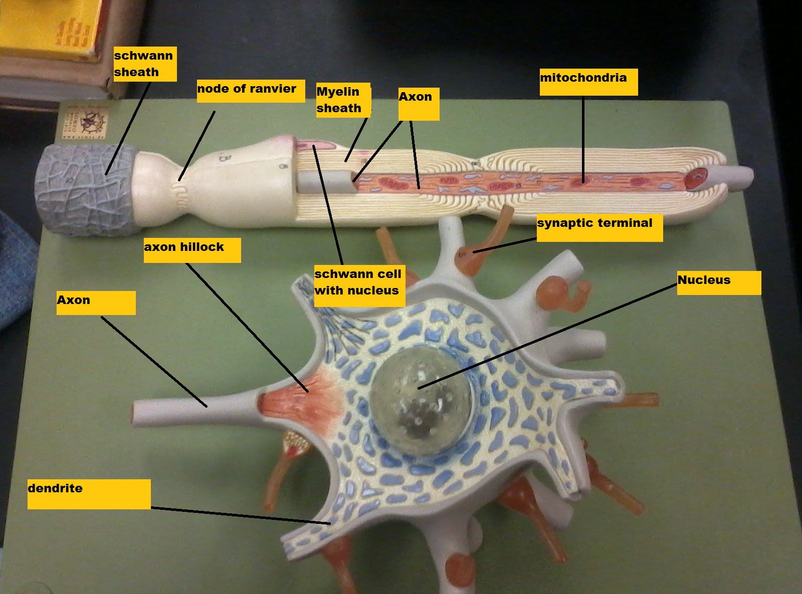
The Hardest Working Label In Hip-Hop: Inside the Rise of Yo Gotti and ... You know CMG's roster by way of rising stars like Moneybagg Yo, EST Gee and 42 Dugg. But for its relentlessly driven leader, the label's rise has been brick-by-brick, and decades in the making. Mnemonics for Heart Anatomy and Physiology (Video) A heart block is an abnormal heart rhythm known as an arrhythmia and can occur anywhere in the specialized conduction system of the heart. The electrical signals telling the heart to contract are partially or totally blocked between the atria and ventricles. Therefore, it is called an atrioventricular, or AV block. Know the Structures and Functions about Your Heart The heart has four chambers: Right atrium Left atrium Right ventricle Left ventricle Atria are smaller than ventricles and have thin, less muscular walls. They are the receiving chambers of the blood, which is delivered to the heart by the large veins. Ventricles are the larger, more muscular pumping chambers that push blood out to the circulation. Histology, Fibroblast - StatPearls - NCBI Bookshelf The ECM is composed primarily of collagen, proteoglycans, and glycoproteins. In homeostasis, the primary collagen type produced by the fibroblast is organ dependent. Type I is predominant in the skin, I and III are prevalent in the interstitium of the lung, liver, and kidney, and IV collagen is abundant in the alveolar basement membrane. [1]
Kidney Structures and Functions Explained (with Picture and Video) The glomerulus connects to a long, convoluted renal tubule which is divided into three functional parts. These consist of the loop of Henle (nephritic loop), the proximal convoluted tubule, and the distal convoluted tubule, which empties into the collecting ducts. These collecting ducts fuse together and enter the papillae of the renal medulla. 14+ Parts Of The Heart With Label | Robhosking Diagram The heart is located in between the two lungs. Anatomy of the human heart. To find a good diagram, go to google images, and type in the internal structure of the human heart. In this interactive, you can label parts of the human heart. 14+ Parts Of The Heart With Label. It pumps blood from the heart to different parts of the body and back to ... The Parts of the Heart and their Functions - Step To Health First, it's fundamental to know that the heart is a hollow organ. It is made up of four cavities: two atria and two ventricles. Also, this organ is divided into a left and a right part. This is because the left atrium is connected to the left ventricle via the mitral valve. Animal Cell: Animal Cell Diagram: Types, and Functions - Embibe Read on to find out its definition, types and structure. Definition of Animal Cell. An animal cell is a eukaryotic cell having membrane-bound cell organelles without a cell wall. The size of the cell varies from a few microns to a few centimetres. For example, the largest animal cell is the ostrich egg measuring 170 mm x 130 mm.
Label every structure on the figure of the heart: image | Study.com
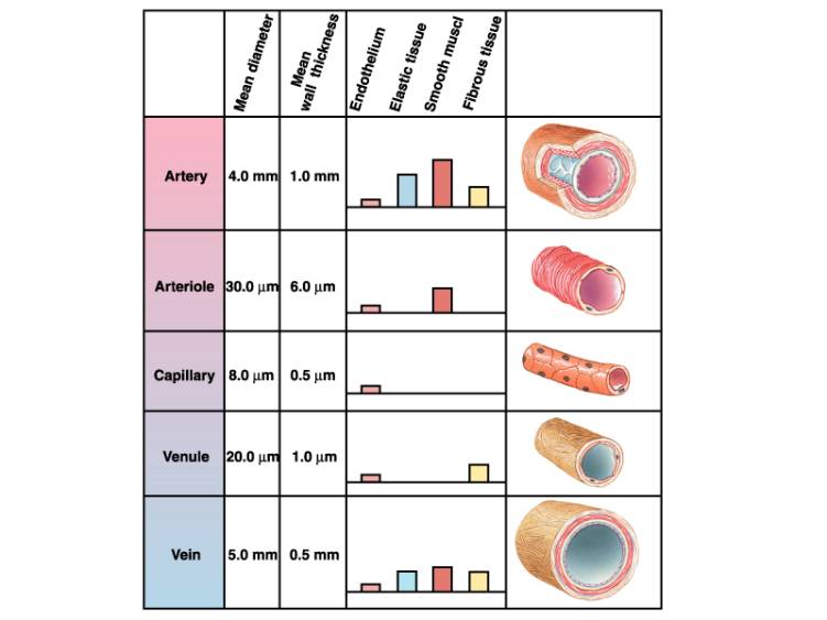
Heart to Heart - Lesson - TeachEngineering The heart itself is composed of muscle tissue, and is subdivided into three layers: Epicardium — this is the outermost layer of the heart, and it helps to reduce friction by containing some fat amongst the coronary arteries. Myocardium — this thick layer of cardiac muscle is the part of the heart that contracts to create a pumping action.
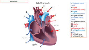
Anatomy Of The Heart Quiz Label
Circulatory System Diagram | New Health Advisor Heart consists of four chambers namely right atria, left atria, right ventricle and left ventricle. Both the atrium and ventricles are separated from each other with a muscular septum. Valves are present in between atria and ventricle which helps in draining the blood from upper part to lower part of the body.

MRI BLOG: Cardiac Anatomy
Path of Blood Through the Heart - New Health Advisor Here are the basic parts of the heart: 1. Right Atrium The heart can be divided into right and left halves, as well as into the upper and lower chambers. There are two upper chambers called atria and two lower chambers called ventricles. On each side of the heart, there are one atrium and one ventricle.

The Heart Diagrams Labeled and Unlabeled
Central nervous system: Anatomy, structure, function | Kenhub Central nervous system anatomy. The central nervous system (CNS) is a division of the nervous system whose function is to analyze and integrate various intra- and extrapersonal information, as well as to generate a coordinated response to these stimuli. Put simply, the CNS is the supreme command center of the body. The CNS consists of two organs which are continuous with each other; the brain ...

Please label the following heart anatomy:
Heart - Wikipedia The wall of the heart is made up of three layers: epicardium, myocardium, and endocardium. The heart pumps blood with a rhythm determined by a group of pacemaker cells in the sinoatrial node. These generate a current that causes the heart to contract, traveling through the atrioventricular node and along the conduction system of the heart.
The Circulatory System
Blood supply to the brain: Anatomy of cerebral arteries | Kenhub The internal carotid artery is one of two branches of the common carotid artery. It is responsible for supplying a large portion of the anterior and middle parts of the brain. A new classification system divides the internal carotid artery into four parts; cervical in the neck, petrous in the base of the skull, cavernous within the cavernous sinus and intracranial above the cavernous sinus.
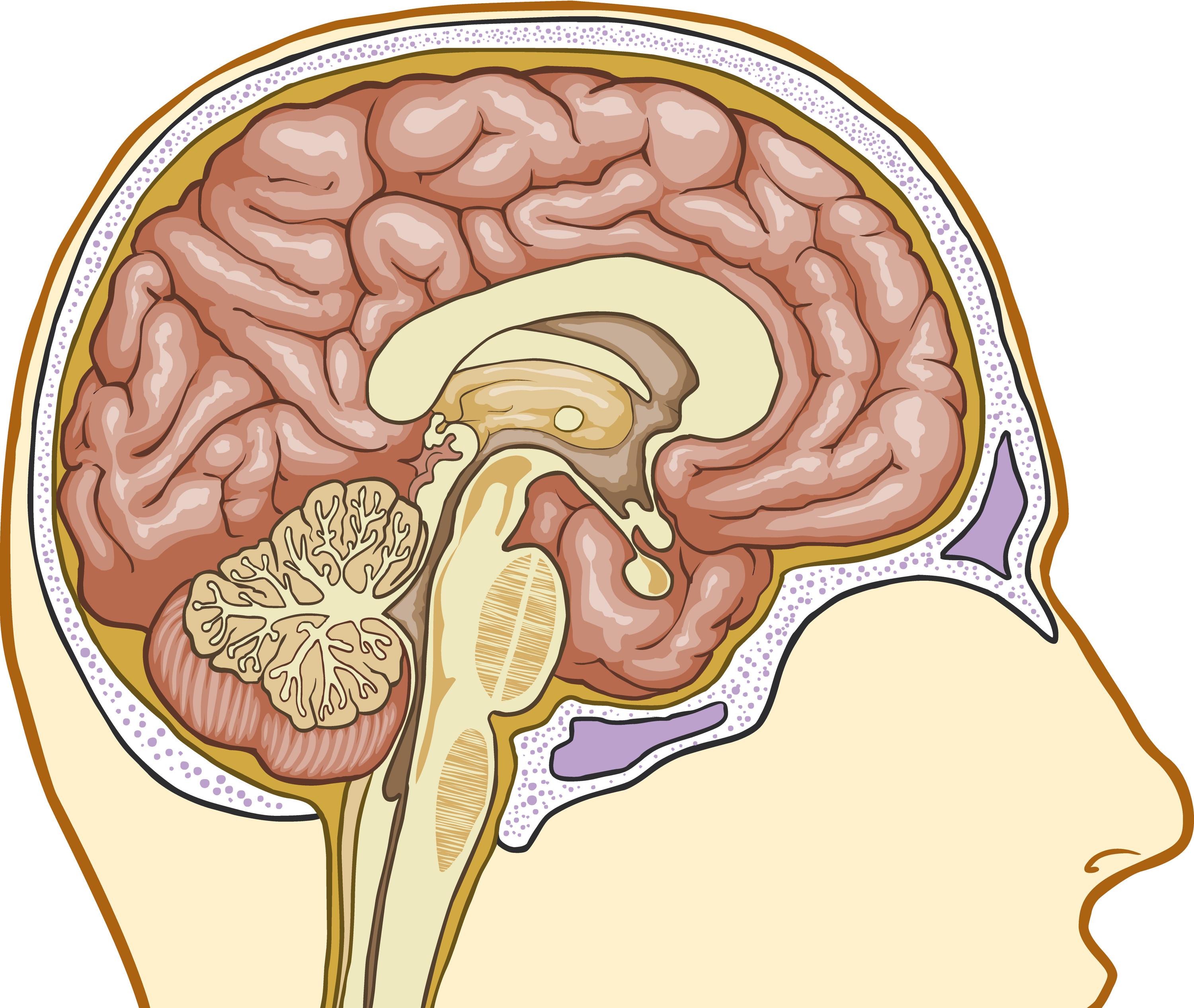
Basic Structure Of The Thalamus - Interactive Biology, with Leslie Samuel
ever wonder why a drawing of a heart - iphonexswallpaperhd But the real heart has a complex structure. The heart is a popular symbol in art doodling and drawing. The heart symbol we use today came from the. How to draw an impossible heart shape. Aristotle the famous philosopher said the human heart had three. The easiest way to draw a heart is with circles. ... Label. 2 35mm 5 8 academy ...

In this diagram they are showing the function of the heart as they have labels to the parts of ...
Internal Anatomy Of The Heart - Heart Failure - GUWS Medical A cross section cut through the heart reveals three layers (Fig. 7): (1) a superficial visceral pericardium or epicardium (epi = "upon" + "heart"); (2) a middle myocardium (myo = "muscle" + "heart"); and (3) a deep lining called the "endocardium" (endo = "within," derived from the endoderm layer of the embryonic trilamina).The endocardium is a sheet of epithelium called endothelium that rests ...

Print Equations flashcards | Easy Notecards
The Cardiac Cycle - Pressures in The Heart - TeachMePhysiology The Cardiac Cycle. At rest, the heart pumps around 5L of blood around the body every minute, but this can increase massively during exercise. To achieve this high output efficiently, the heart works through a carefully controlled sequence with every heartbeat - this sequence of events is known as the cardiac cycle.

Heart label diagram
human heart drawing labeled The heart is made up of four chambers. The upper two chambers of the heart are called auricles. I havent looked through all of the structures but your superior vena cava is incorrectly labeled at the ascending side of the aortic. To make the superior vena cava draw a tube coming from the top of the right atrium.

Human Heart Labeling
Morphine - StatPearls - NCBI Bookshelf FDA-approved usage of morphine sulfate includes moderate to severe pain that may be acute or chronic. Most commonly used in pain management, morphine provides major relief to patients afflicted with pain. Clinical situations that benefit greatly by medicating with morphine include management of palliative/end-of-life care, active cancer treatment, and vaso-occlusive pain during sickle cell ...
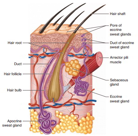
Epidermis And Accessory Structure Formed By The Epidermis And Their Functions
Thalamus | Anatomy, Location, Structure, Function & Physiology Structure Thalamus is an egg-shaped mass of grey matter. It also contains white matter to some extent. White Matter The white matter of thalamus consists of three laminae. These are mentioned below. On the superior surface, thalamus is covered by a thin layer of white matter. This layer of white matter is called stratum zonale.
Chapter 14 Nervous Tissue - Biology 4 Human AnatomyProfessor Julie GallagherBarstow Community ...
Circulatory system - Wikipedia The circulatory system includes the heart, blood vessels, and blood. The cardiovascular system in all vertebrates, consists of the heart and blood vessels. The circulatory system is further divided into two major circuits - a pulmonary circulation, and a systemic circulation. The pulmonary circulation is a circuit loop from the right heart taking deoxygenated blood to the lungs where it is ...

Dollhouse miniature Halloween | Dollhouse halloween, Halloween miniatures, Halloween shadow box
Histology, Lung - StatPearls - NCBI Bookshelf The respiratory portion of the lung consists of respiratory bronchiole, alveolar duct, alveolar sac, and finally alveoli where actual respiration takes place. In the conducting zone, the air is moistened, warmed, and filtered before it reaches the start of the respiratory region at the respiratory bronchioles.
Related Items

Kids' Health - Topics - Your heart | Heart for kids, Heart diagram, Heart lesson
Post a Comment for "45 heart structure and labels"