44 diagram of the lungs with labels
Labeled diagram of the lungs/respiratory system. - SERC View Original Image at Full Size. Labeled diagram of the lungs/respiratory system. Image 37789 is a 1125 by 1408 pixel PNG Uploaded: Jan10 14. Last Modified: 2014-01-10 12:15:34 Diagram of Human Heart and Blood Circulation in It Four Chambers of the Heart and Blood Circulation. The shape of the human heart is like an upside-down pear, weighing between 7-15 ounces, and is little larger than the size of the fist. It is located between the lungs, in the middle of the chest, behind and slightly to the left of the breast bone. The heart, one of the most significant organs ...
Label the lung diagram - Quizlet Start studying Label the lung diagram. Learn vocabulary, terms, and more with flashcards, games, and other study tools.

Diagram of the lungs with labels
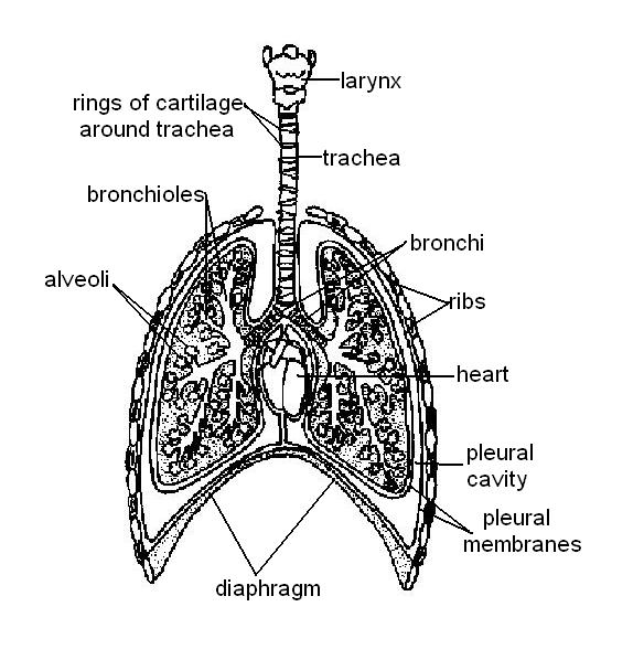
Heart Anatomy: Labeled Diagram, Structures, Function, and Blood Flow Function and anatomy of the heart made easy using labeled diagrams of cardiac structures and blood flow through the atria, ventricles, valves, aorta, pulmonary arteries veins, superior inferior vena cava, and chambers. ... Trick 3: You can use the lobes of the lungs as a reference. The RIGHT lung has 3 lobes, and the TRIcuspid valve (3) ... Lungs (Human Anatomy): Picture, Function, Definition, Conditions - WebMD Next. The lungs are a pair of spongy, air-filled organs located on either side of the chest (thorax). The trachea (windpipe) conducts inhaled air into the lungs through its tubular branches ... Label Lungs Diagram Printout - EnchantedLearning.com Human Anatomy. Read the definitions below, then label the lung anatomy diagram. bronchial tree - the system of airways within the lungs, which bring air from the trachea to the lung's tiny air sacs (alveoli). cardiac notch - the indentation in the left lung that provides room for the heart. diaphragm - a muscular membrane under the lungs.
Diagram of the lungs with labels. Label Lungs Diagram Printout - EnchantedLearning.com bronchial tree - the system of airways within the lungs, which bring air from the trachea to the lung's tiny air sacs (alveoli). cardiac notch - the indentation in the left lung that provides room for the heart. diaphragm - a muscular membrane under the lungs. larynx - a muscular structure at the top of the trachea, containing the vocal cords. left inferior lobe - the bottom lobe of the lung ... Lungs Diagram High Resolution Stock Photography and Images - Alamy Lungs Labeled Diagram. Woman anatomy cardiovascular system with skeleton and internal organs, rear and front views. Lungs and human respiration. Anatomy graphic, Illustration. Smoking education concept - body, health, healthy living, medicine, medical ... The Lungs - Labelled diagram - Wordwall Drag and drop the pins to their correct place on the image.. trachea, bronchi, bronchioles, Alveoli, Heart, Diaphragm , Rib, Intercostal Muscle . Diagram Lungs Stock Illustrations - 2,491 Diagram Lungs ... - Dreamstime Download 2,491 Diagram Lungs Stock Illustrations, Vectors & Clipart for FREE or amazingly low rates! New users enjoy 60% OFF. 186,048,303 stock photos online. ... Labeled diagram with brain sections. Cranial nerves vector illustration. Labeled diagram with brain sections and its. Different systems of human body diagram. Illustration.
Respiratory System Labeling Interactive Quiz About this Quiz. This is an online quiz called Respiratory System Labeling Interactive. There is a printable worksheet available for download here so you can take the quiz with pen and paper. Lung Diagram Labeled | EdrawMax Template The following is the elaborated diagram of human lungs. In the following lung labeled diagram, we have shown Thyroid cartilage, Cricoid Cartilage, Tracheal Cartilage, Apex, Left Upper Lobe, Hilum, Left Bronchus, Oblique Fissure, Bronchioles, Left Lower lobe, Base of lung, cardiac notch, right lower lobe, oblique fissure, right middle lobe, horizontal fissure, right bronchus, right upper lobe ... Lungs: Definition, Location, Anatomy, Function, Diagram, Diseases Where are the Lungs Located. The lungs are located a little toward the posterior part of the human body, just below the collarbone, extending down to the diaphragm, the muscular partition that separates the chest and abdominal cavities.The left and right lungs are situated on the two sides of the body with the heart, another vital organ in the thoracic cavity, located a little in front of, and ... Lung Anatomy, Function, and Diagrams - Healthline The lungs begin at the bottom of your trachea (windpipe). The trachea is a tube that carries the air in and out of your lungs. Each lung has a tube called a bronchus that connects to the trachea ...
Fully Labelled Diagram Alveolus Lungs Showing Stock ... - Shutterstock Fully labelled diagram of the alveolus in the lungs showing gaseous exchange. Formats. EPS. 1114 × 800 pixels • 3.7 × 2.7 in • DPI 300 • JPG. Show more. Contributor. S. Steve Cymro. Find Fully Labelled Diagram Alveolus Lungs Showing stock images in HD and millions of other royalty-free stock photos, illustrations and vectors in the ... Lungs Diagram Labeled Pictures, Images and Stock Photos Search from Lungs Diagram Labeled stock photos, pictures and royalty-free images from iStock. Find high-quality stock photos that you won't find anywhere else. Respiratory System Anatomy, Diagram & Function | Healthline Respiratory. The respiratory system, which includes air passages, pulmonary vessels, the lungs, and breathing muscles, aids the body in the exchange of gases between the air and blood, and between ... Diagram Of The Respiratory System With Labels Stock Photos, Pictures ... Lungs with Alveoli Labeled CG image of woman's chest area showing both lungs in isolation, with magnified view of alveoli air sacs labeled on faded flesh tone and white. diagram of the respiratory system with labels stock pictures, royalty-free photos & images
Lung anatomy | Medical library, Lung anatomy, Human anatomy and physiology
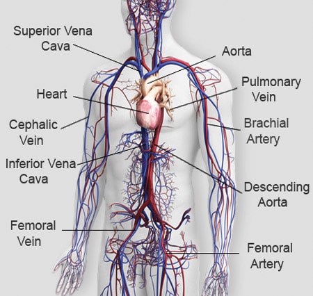
Label Lungs Diagram Printout - EnchantedLearning.com | Respiratory ... Diagram illustrating the paths of blood flow through the heart. Blood enters the heart through two large veins, the inferior and superior vena cava, emptying oxygen-poor blood from the body into the right atrium.

Circulatory System Diagram - Cardiovascular System and Blood Circulation Diagram
› stock-photo › female-anatomy-diagramFemale Anatomy Diagram Stock Photos and Images - Alamy Find the perfect female anatomy diagram stock photo. Huge collection, amazing choice, 100+ million high quality, affordable RF and RM images. No need to register, buy now!
Respiration Worksheet Answers - WikiEducator
Lung Diagram | Free Lung Diagram Template - Edrawsoft The lung diagram template here clearly presents a pair of spongy on both side of the chest. Simply hitting on the template to learn more parts including pleura, ribs, bronchi, alveoli and more. Feel free to find out more human anatomy templates and symbols in the free download version. Lab Apparatus List. 64705.

Abdominal Anatomy Male - Human Body Diagram Without Labels , Transparent Cartoon, Free Cliparts ...
› science › articleGenome-wide CRISPR Screen in a Mouse Model of Tumor Growth ... Mar 12, 2015 · We derived and cloned a cell line (Chen et al., 2014) from a mouse non-small-cell lung cancer (NSCLC) (Kumar et al., 2009).This cell line possesses an oncogenic Kras in conjunction with homozygous p53 and heterozygous Dicer1 loss of function (Kras G12D/+;p53 −/−;Dicer1 +/−, denoted KPD) and is capable of inducing tumors when transplanted into immunocompromised mice (Chen et al., 2014 ...

Respiratory system - wikidoc
DIAGRAMS: Lungs Cross Section - Labeled - Body Diagram | Body diagram ... The Human Body - Explore a working model of the body. Every part is animated and interactive: the heart beats, guts gurgle, lungs breathe, the skin feels, and eyes see. Designed for kids ages 4+ to discover what we're made of and how we work. luckyme001. L.

Health Articles: Protect Your 5 Vital Parts of Body
Labeled Diagram of the Human Lungs - Bodytomy Given below is a labeled diagram of the human lungs followed by a brief account of the different parts of the lungs and their functions. Each lung is enclosed inside a sac called pleura, which is a double-membrane structure formed by a smooth membrane called serous membrane. The outer membrane of this structure is called parietal pleura and is ...

Circulatory system
Lobes of the Lung - SmartDraw Lobes of the Lung. Create healthcare diagrams like this example called Lobes of the Lung in minutes with SmartDraw. SmartDraw includes 1000s of professional healthcare and anatomy chart templates that you can modify and make your own.
Your Lungs & Respiratory System
lisbdnet.com › diagram-of-how-blood-flows-throughdiagram of how blood flows through the heart – Lisbdnet.com Nov 28, 2021 · Your heart first pumps blood to your lungs. Here, the blood picks up oxygen from the air that you have breathed in. The blood (carrying oxygen) then travels back to your heart. The heart gives the blood a second push. How the heart pumps blood ks3? The right ventricle pumps the low-oxygen blood to the lungs to pick up a fresh supply of oxygen ...

Lungs - Anatomy QA
Lungs label - Teaching resources 3728 results for 'lungs label'. Lungs Labelled diagram. by Rbowerkail. KS4 PE. The Lungs Labelled diagram. by Fayeroberts. KS4 Y10 Biology. Lungs Diagram Labelled diagram. by Jon9.
Normal Lung Anatomy
Anatomy of the Lung | SEER Training Anatomy of the Lung. The lungs are the major organs of the respiratory system, and are divided into sections, or lobes.The right lung has three lobes and is slightly larger than the left lung, which has two lobes.. The lungs are separated by the mediastinum.This area contains the heart, trachea, esophagus, and many lymph nodes. The lungs are covered by a protective membrane known as the pleura ...
The circulatory system of mammals - Free ZIMSEC & Cambridge Revision Notes
The Respiratory System (Label Diagram) - ScienceQuiz.net Match each pair by dragging from right to left. When complete click Check button.

More Lung Anatomy
Can you label the lungs? Quiz - PurposeGames.com This is an online quiz called Can you label the lungs? There is a printable worksheet available for download here so you can take the quiz with pen and paper. From the quiz author. Labeling the lungs. This quiz has tags. Click on the tags below to find other quizzes on the same subject. lungs. respiratory system.

Science Biology: S2 Lung dissection
Label Diagram Of The Lungs - Solved Labeling Diagram Directions Label ... Label lungs anatomy diagram printout. It also includes an incredible ar feature! Lung cancer is a leading cause of death in the u.s. Give the diagram a heading and . There are 2 different types of diagram to choose from for students to label. Learn what you need to know about lung cancer. Labeled diagram of the lungs/respiratory system.:
Body Systems
Label the Lungs Diagram | Quizlet Start studying Label the Lungs. Learn vocabulary, terms, and more with flashcards, games, and other study tools.
Post a Comment for "44 diagram of the lungs with labels"